Inside View
A photo essay of brain structures and neural mechanisms
- 8 minute read
- Images

A cross-section of the mouse nasal epithelium
The brain and its functioning are endlessly fascinating to many. So, too, are images of the brain. In this photo essay, we present images captured by Harvard scientists as they conduct research they hope will help unravel neural mysteries.
Their work involves deciphering the connections between regions of the brain, decoding the mechanisms that translate sensory signals from external stimuli, creating maps of the brain’s molecular activity, and investigating the genetics of nervous system tumors. The knowledge they gather further shapes what is known about how the brain oversees our physiological functioning, directs our behavior, becomes diseased, and responds to novel therapeutics.
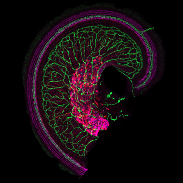
Photo by: Katelyn Comeau, PhD candidate
PI: Lisa Goodrich, HMS professor of neurobiology
The apex of the snail-shaped cochlea, our hearing organ, is shown above with its dense vascular network (green). The hair cells (magenta), which detect sound, are innervated by the primary auditory neurons, whose cell bodies and axons are also in magenta.
The neurons and sensory cells of the mammalian cochlea relay to the brain information on the diverse sounds that surround us daily, forming the basis of our sense of hearing. The auditory system is complicated, and researchers in the field continue to work to answer fundamental neurobiological questions. Past work in Goodrich’s lab has shed light on the development and molecular diversity of primary auditory neurons. Current work by Comeau builds on this earlier research by studying newly identified molecules that help neurons make the proper synaptic connections with the sensory cells in the cochlea.
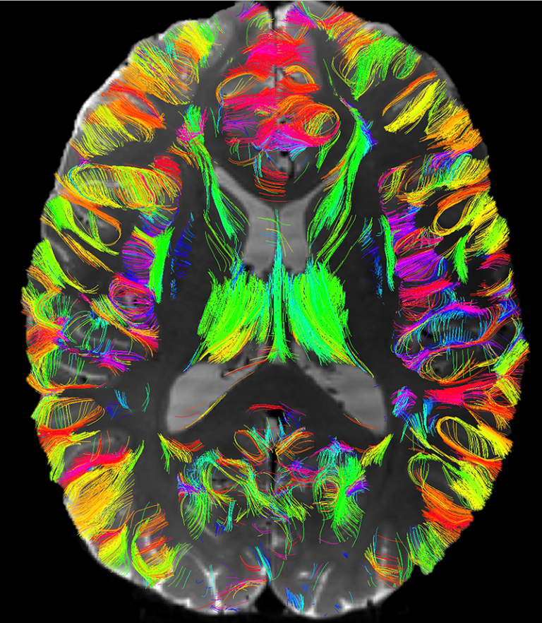
Photo by: Fan Zhang, former HMS instructor in radiology, Brigham and Women’s Hospital
PIs: Nikolaos Makris, HMS professor of psychiatry, Massachusetts General Hospital
Lauren Jean O’Donnell, HMS associate professor of radiology, Brigham and Women’s
Yogesh Rathi, HMS associate professor of psychiatry, Brigham and Women’s
Lab: Lauren Jean O’Donnell
The superficial white matter of the human brain is located beneath the cortex and plays an important role in cortico-cortical communication. The photo above was captured using ultrahigh-resolution diffusion magnetic resonance imaging and shows previously invisible u-shaped connections of the superficial white matter. The colors indicate the overall 3D orientation of fibers in the brain and their estimated white matter connections.
The O’Donnell lab is working to create the first atlas of the human brain’s superficial white matter using submillimeter ultrahigh-resolution diffusion MRI. Located between the deep white matter and the cortex, the superficial white matter plays an important role in neurodevelopment and aging and has been implicated in a large number of diseases, yet is vastly underrepresented in current descriptions of the human brain connectome. The creation of comprehensive, anatomically curated maps of the superficial white matter would allow it to be studied in health and disease.
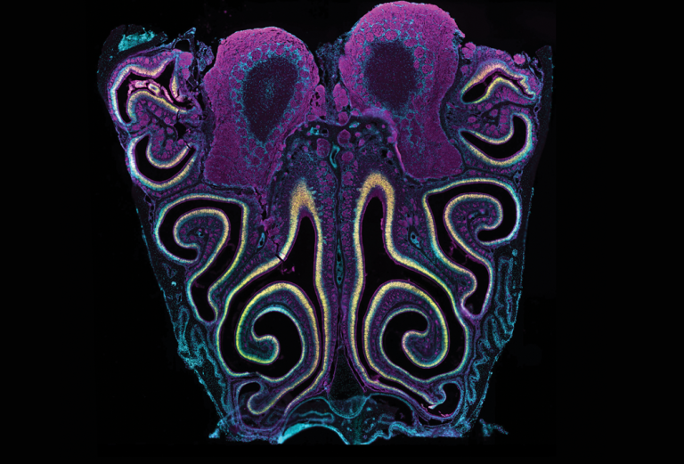
Photo by: David Brann, PhD candidate
PI: Sandeep Robert Datta, MD ’04 PhD ’04, HMS professor of neurobiology
A cross-section of the mouse nasal epithelium is shown above. Olfactory sensory neurons (yellow) detect volatile odorants and relay the information via their axons (magenta) directly to the brain, where the axons coalesce into structures known as glomeruli in the olfactory bulb, the rounded structures at the top. Cell nuclei (cyan) are stained with an agent known as DAPI.
Nasal sensory neurons detect odors via a diverse set of receptors — the largest gene family in our genome. The Datta lab is developing high-throughput sequencing approaches to identify which odors activate which receptors: Each odor interacts with a subset of receptors and each receptor interacts with a subset of odors. The aim is to decipher the molecular logic that underpins our sense of smell.
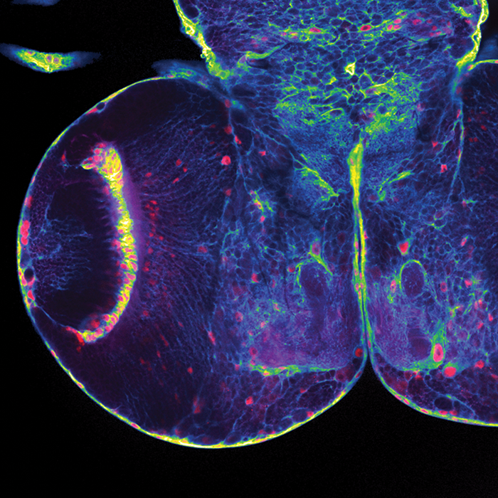
Photo by: Torrey Mandigo, postdoctoral fellow
PI: James Walker, HMS assistant professor of neurology in the Center for Genomic Medicine, Massachusetts General Hospital
The fruit fly larval brain shown in the above image is used by researchers to study glioma, a type of brain tumor. The different types of brain cells shown include glial cells (blue), which support, nourish, and protect neurons, glial nuclei (red), and neuropils (green), which are dense networks of fine glial processes and neuronal processes (axons and dendrites).
Neurofibromatosis type 1 is a genetic disease characterized by tumors of the nervous system. The Walker lab has developed fruit fly models of the disorder and uses them to improve our understanding of the mechanism that drives neurofibromatosis type 1, with the goal of identifying potential therapeutic targets.
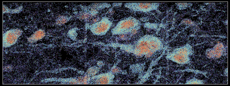
Photo by: Rongxin Fang, postdoctoral fellow
PI: Xiaowei Zhuang, Harvard University professor of chemistry and chemical biology and professor of physics
Multiplexed RNA imaging of part of the human brain was used to produce the above image, which shows RNA molecules (various colors) expressed from 4,000 different genes in individual cells. The imaging technique was used to identify transcriptionally distinct cell populations and to generate a molecularly defined and spatially resolved cell atlas of the human cortices.
The Zhuang lab develops novel imaging methods, molecular probes, and image analysis algorithms, and uses these tools to study a variety of biological problems. The research that produced this image involved the development of high-resolution, spatially resolved single-cell imaging methods that can be used to study how the spatial and molecular architecture of the mammalian brain alters during evolution and in diseases.
The images presented in this essay won special recognition in the annual The Beauty of the Brain photo contest from the Harvard Brain Science Initiative, a cross-schools effort launched in 2014 by the Office of the Provost at Harvard University. The initiative is jointly directed by David Ginty, the Edward R. and Anne G. Lefler Professor of Neurobiology at HMS and chair of the Department of Neurobiology in the Blavatnik Institute at HMS, and Venkatesh Murthy, the Raymond Leo Erikson Life Sciences Professor of Molecular and Cellular Biology at Harvard University and director of the Harvard Center for Brain Science.