What Happens to the Brain Under Anesthesia
When nobody could explain exactly how anesthesia renders a person unconscious, Emery Brown went looking for answers
- 8 minute read
- Interview
Photo: Matt Kalinowski

Photo: Matt Kalinowski
On October 16, 1846, physicians filled the surgical amphitheater at Massachusetts General Hospital to watch dentist William Morton test a bold new idea.
Morton held a glass inhaler filled with an ether-soaked sponge and asked a patient, Gilbert Abbott, to breathe in. Within minutes, Abbott lay motionless. Surgeon John Collins Warren then made an incision in Abbott’s neck and removed a tumor. When Abbott awoke, he reported having felt no pain. It was the first public demonstration of the use of ether for surgical anesthesia, and a moment that changed medical history.
About 140 years later, in the same hospital, medical student Emery Brown, MD ’87, started his rotation in anesthesiology. He was quickly hooked. He loved channeling his knowledge of physiology and pharmacology to manage a patient moment-by-moment in the operating room. But there was one problem that began to bug him: Although general anesthesia had been used millions of times since 1846, nobody in the field seemed to really know — or even care — how the practice actually worked.
“If the anesthetic drugs are acting in the brain and we know the brain’s circuits,” wondered Brown, who is now the HMS Warren M. Zapol Professor of Anaesthesia at Mass General, “can’t we start to think about where and how they’re acting in the brain?”
Brown’s insistence on that question may have stemmed from his unique path. The son of high school math teachers in Ocala, Florida, he studied applied mathematics at Harvard before pursuing an MD-PhD. Earning his PhD in statistics — a highly unusual path for a medical trainee — he developed statistical methods to understand neuroscience data. He didn’t know it then, but that expertise would come in handy once those nagging questions about the neuroscience of anesthesia finally felt too big to ignore.
After publishing his critiques of anesthesiology in a 2010 article in the New England Journal of Medicine and receiving an NIH Pioneer Award to conduct what the agency calls “high-risk, high-reward research,” he started using electroencephalography, or EEG, to record and analyze brain wave patterns of patients under general anesthesia.
It was like having a secret handshake, and only those who knew the handshake could administer anesthesia.
Brown went on to demonstrate that anesthesia is nothing like natural sleep; instead, it’s more like a coma, with large, slow oscillations in neural activity that prevent different parts of the brain from communicating. He has shown that different classes of anesthetic drugs create unique EEG patterns and that those patterns differ depending on dosage and a patient’s age and health status. His research has moved the field toward a more rigorous, brain-centered understanding of anesthesia and of unconsciousness itself.
Brown is the first anesthesiologist, the first statistician, and the first African American to be elected to all three of the National Academies of Sciences, Engineering, and Medicine. He also received the 2024 National Medal of Science. In addition to his role at HMS, Brown is the Edward Hood Taplin Professor of Medical Engineering and Computational Neuroscience at the Massachusetts Institute of Technology.
Harvard Medicine associate editor Molly McDonough spoke with Brown to learn more about what we now know — and still don’t — about anesthesia and the brain. The interview has been edited for length and clarity.
There was a long-standing idea that anesthesiologists could use anesthesia without really understanding how it worked. Why didn’t that sit right with you?
I think anesthesiologists enjoyed being able to say, “We do it, but we don’t know how it works.” It was like having a secret handshake, and only those who knew the handshake could administer anesthesia. But there’s no way you can improve it if you have no idea how it works.
There were some obvious phenomena that I could see were occurring based on neuroscience. For example, when a neurologist is evaluating a patient in coma, they often check the patient’s oculocephalic reflex — how the eyes move — not because they’re interested in the eyes, but because that gives them a window into the part of the brainstem where the circuits controlling arousal are located. And if you check that reflex in someone under general anesthesia, you see that the reflex is lost. Also, a patient would need to lose physical sensation in order to tolerate a surgical procedure. It seemed reasonable to begin thinking of general anesthesia as a pharmacologically mediated, reversible coma.
You’ve pointed out that anesthesiologists tend to use the word “sleep” in the context of general anesthesia, which doesn’t really make sense.
Anesthesiologists tell patients, “You’re going to go to sleep.” And then they say to each other, “Well, I don’t really mean that.” But what do they mean? They say sleep because, understandably, they don’t want to frighten patients by telling them they will be in a pharmacologically mediated, reversible coma.
I realized that anesthesiologists didn’t have a clear definition of general anesthesia. So, I stated one. There are four components: You’re unconscious, you don’t feel pain, you don’t form memories, and you’re not moving around. The key point is to create these states while keeping the patient physiologically stable and bringing them out once the surgery is finished. That started to clarify my thinking; it allowed me to think about the various components and how we were generating each of them.
It also changed how I spoke to patients. I won’t say, “I’m going to put you in a pharmacologically mediated, reversible coma, but don’t worry, I can wake you up.” But I can say, “Look, you’re going to be unconscious. You won’t realize anything that’s going on or perceive any pain. You won’t form any memories of what’s happening. I’ll probably give you some medications to relax your muscles to make it easier for the surgeons to operate. I’ll be with you the whole time, watching your heart rate, blood pressure, blood oxygen levels, carbon dioxide levels, and temperature to make sure they are stable. When all is done, the muscle relaxant has been reversed, and the effects of the other anesthetics have worn off, I’ll wake you up.” So, I just give them the definition of general anesthesia in lay terms.
Do we understand now how anesthesia works?
I think we now understand the principal mechanisms through which it works, particularly for widely used agents like propofol and the ether anesthetics — sevoflurane, isoflurane, and desflurane.
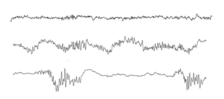
In 2011, I started using the EEG to monitor all my patients during general anesthesia. It’s very impressive to watch: As you administer the medications, you see the patients’ brain wave oscillations start to change. During the normal eyes open, awake state, EEG oscillations have frequencies of around 40 cycles per second with small amplitudes. As the anesthetic quickly reaches various parts of the brain, the oscillations become larger in amplitude and decrease in frequency — dropping to about 8 to 14 cycles per second (called alpha oscillations) and 0.3 to 4 cycles per second (called slow-delta oscillations). At the same time, individual neurons fire far less often, slowing from about 10 to 12 times per second to roughly once every one to two seconds.
Hence, one of the things that propofol and the ether anesthetics are doing is dramatically altering dynamics so that normal communication among brain regions can’t continue. Because these oscillations are not natural and are sustained sometimes for several hours, they explain why postoperative brain dysfunction can be common, particularly in the elderly.
Another thing we’ve learned is that high doses of these anesthetics alter the brain’s ability to produce ATP, the primary energy source for cells. This manifests in a specific pattern on the EEG called “burst suppression,” a warning sign that the brain’s metabolism is being altered. We see this commonly in older patients, 60 years and over. They enter this state very readily with small doses of these anesthetics. If we are not monitoring, burst suppression is not detected, and patients are overdosed.
But that continuous EEG monitoring is not standard, right?
Unfortunately, that’s right. EEG monitoring is used much more regularly in Europe, South America, and perhaps in Asia, but not here in the United States. I think this is a fundamental shortcoming. There are monitors that take EEG brain wave data and simplify it into a single number or index displayed on a screen. Anesthesiologists are basically told, “Don’t worry about the unprocessed EEG signal, just use the index. As long as it’s within a certain range, the patient is fine.” It’s a total black box. And there are well-known situations in which the indices are frequently incorrect, such as with children, and with certain anesthetics like ketamine and dexmedetomidine.
Within weeks of when I first started reading EEG patterns directly, I could see unique signatures depending on which class of drugs we were using and that those signatures also changed depending on how old the patients were and their state of health. It was obvious, but no one had sat down and looked at it.
So if you’re just looking at a number and not the actual EEG signatures, the risk is that you might assume someone is unconscious when they actually have awareness?
Actually, the more common scenario is that if you’re anesthetizing someone and you’re worried about them being awake, your natural action is to give them more anesthesia. So you tend to overdose patients because you do not completely trust the index. People being awake under anesthesia is a horrible occurrence. However, it is very uncommon. What’s more common is that patients wake up with delirium or brain dysfunction because they had too much anesthesia.
Is that the issue you’re trying to address in your research now?
Two things. One is teaching anesthesiologists to read real-time EEG, making use of what we already know. The second is to build a closed-loop system to deliver anesthesia. It’s like putting a plane on autopilot, reading the EEG in real time and using it to dose the anesthetic to maintain a certain level of unconsciousness. We have good reasons to believe this works based on animal studies we’ve recently completed.
By thinking of anesthesia as a measurable form of unconsciousness, does that tell us anything about what consciousness actually is?
When people ask me what I study, I say that I study unconsciousness, because that’s what I want to create in the most sound way for the purpose of anesthesia care. But we know that a person can be in a coma because of a stroke that affects the cortex, the brainstem, or the thalamus. There are all sorts of ways of turning the system off. That suggests the conscious process depends on these brain areas being well integrated and communicating in such a way that you’re awake and you’re aware.
To the extent that I can map how different approaches to anesthesia diminish awareness, alter perception, or produce unconsciousness, I can provide information to investigators who study the harder problem: How does the brain integrate information to create consciousness? We don’t need to wait until that problem is solved, though, to improve anesthesia care. I think that well before we figure out all the details of consciousness, we can develop more sound, neurophysiology-based approaches to anesthesia.
If you could travel back in time and explain the modern science of anesthesia to William Morton and John Collins Warren, what do you think would surprise them most?
I think they’d be surprised to know that a large fraction, maybe 70 percent, of the way anesthesia is practiced today is essentially the way they did it back then. We still widely use ethers that patients breathe while they’re having surgery.
It’s really interesting: Before the use of ether for anesthesia was discovered, do you know what the primary area of research was in surgery? It was coming up with better ways to strap patients down. The discovery of ether changed surgery overnight: from butchery and intolerable pain to more humane therapeutic procedures. It has to be acknowledged as one of medicine’s greatest innovations.
So I’d say, “Dr. Morton, you made a truly transformative discovery that we’re still using today, almost the same way. The world is grateful for this. It inspires us to use our deeper understanding of neuroscience and the brain to create the next-generation solution.”
Molly McDonough is the associate editor of Harvard Medicine.