
The neurobiology behind our sense of touch is fascinating and complicated. It also can be visually stunning.
For two decades, researchers in the laboratory of David Ginty, the Edward R. and Anne G. Lefler Professor of Neurobiology in the Blavatnik Institute at HMS, have investigated the development, organization, and function of the neurons and neural circuits that underlie our sense of touch. Along the way, they have captured breathtaking images of cutaneous sensory neurons in action, illuminating how touch allows us to perceive and respond to our world.
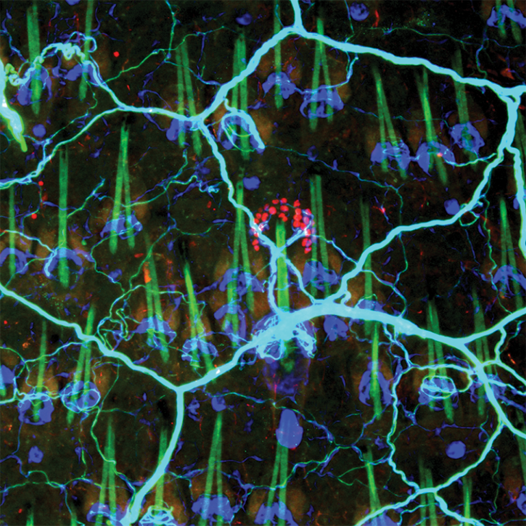
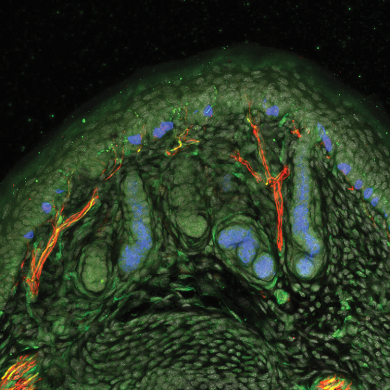
The blue-green processes of sensory neurons form a network of nerve endings in mouse skin. Some of these endings associate with touch-sensitive skin cells (red). The green tent-like structures represent hair shafts. Photo: Michael Rutlin
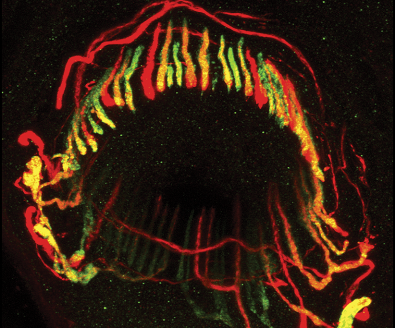
The red, green, and yellow colors reveal the intermingled endings of three sensory neurons found around a mouse hair follicle. This type of neuronal ending, called a lanceolate ending, responds to minute deflections of hair by sending electrical signals to the spinal cord, alerting the animal to the movement. Lanceolate endings associated with hair follicles have been observed in all mammals studied thus far. Photo: Lishi Li
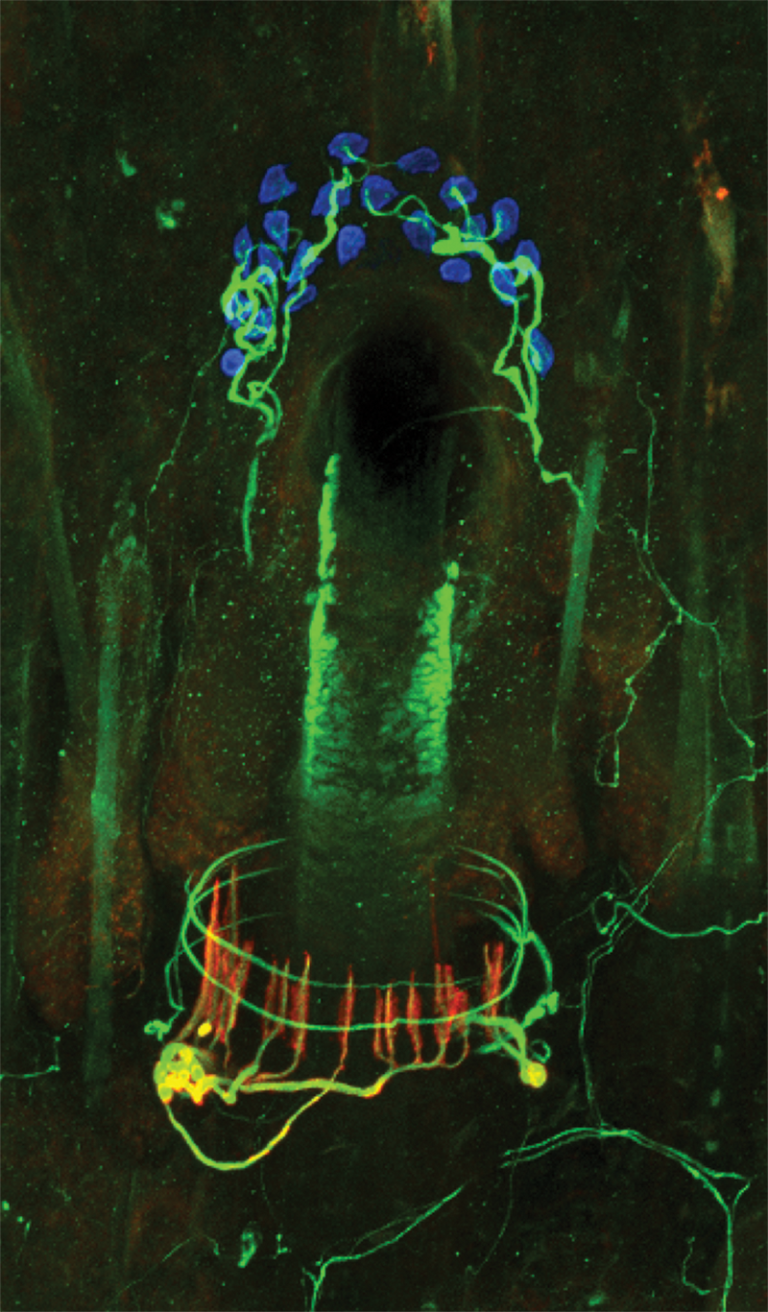
This follicle of a mouse hair shows the endings of three types of sensory neurons, each capable of detecting changes affecting a hair shaft and surrounding skin. The longitudinal lanceolate endings (red) allow the neuron to respond to hair deflections while the neurons that travel the circumference of the follicle (green) respond to gentle stroking of the skin. The third type of neuron (also green) associates with specialized skin cells known as Merkle cells (blue discs). These neurons can respond repeatedly to indentation of the skin. Photo: Victoria Abraira
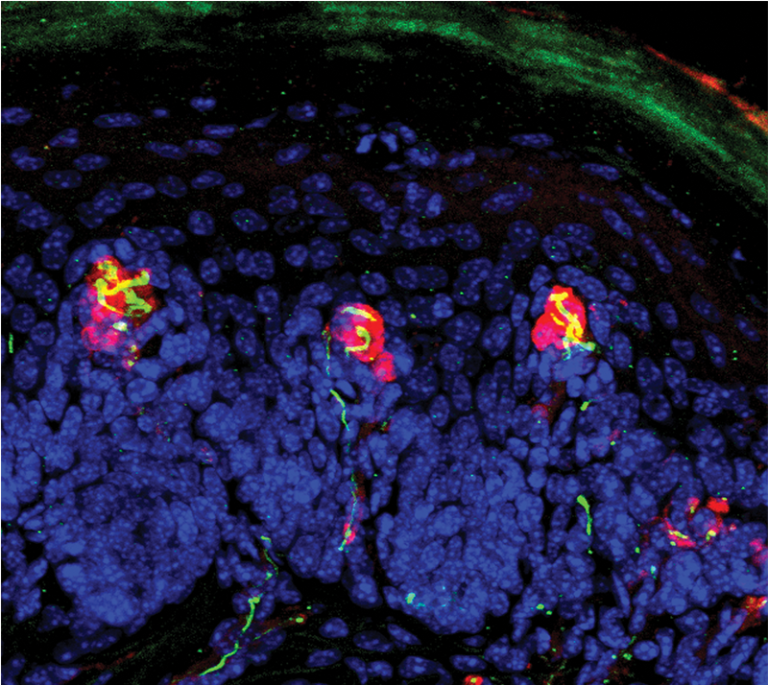
These skin cells (blue) and sensory neuronal endings (yellow) that terminate in close association with nonneuronal lamellar cells (red) make up mechanosensory end organs known as Meissner corpuscles. Highly sensitive to skin indentation, vibration, and gentle movement across the skin, Meissner corpuscles are found in the hairless skin of our fingertips. Photo: Yin Liu
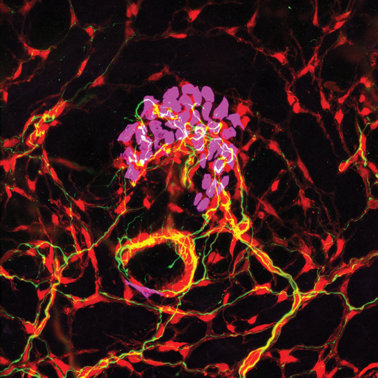
Touch-sensitive neurons form specialized endings in the skin that are critical to our response to any light touch stimuli we might encounter. The touch-sensitive neurons, shown in green or white, are associated with ensheathing support cells (red) and a group of touch-sensitive skin cells (purple) that are located around the mouth of a hair follicle. The green neurons that form circular structures with small spiking processes around a follicle respond to hair deflection. The neurons that form endings associated with the touch-sensitive skin cells respond to skin indentation. Photo: Katelyn Comeau and Shan Meltzer
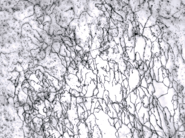
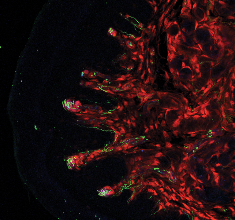
This image shows a single-labeled neuron that forms complex free nerve endings in hairy skin. Photo: Celine Santiago
This confocal microscope image shows sensory nerve endings (green and red) found in the hairless skin of palms, soles of feet, and fingertips. Sweat glands and specialized touch cells are shown in blue. The neuronal cells that send these endings to the skin detect light touch and can help in distinguishing textures and in sensing indentation to and movement on the skin’s surface. Photo: Charalampia Koutsioumpa
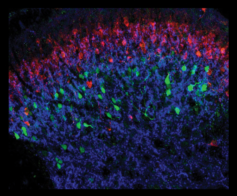
The interneurons highlighted in red and green are two of more than a dozen distinct types located in the dorsal region of the spinal cord. These interneurons receive direct input from several of the sensory neuron types that project to the skin. Photo: Victoria Abraira
A section of non-hairy skin from the forepaw of a mouse showing the neurons that innervate the toe pad (green). A subset of glial cells that are closely associated with the axonal endings of the neurons (red) form around corpuscles in the toe pad. Photo: Celine Santiago