The Birth of Parallel Imaging
How a doodle on a napkin changed medical imaging and launched a young doctor’s career
- 5 minute read
- Perspective

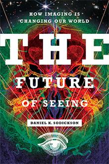
Daniel Sodickson presented his research on simultaneous acquisition of spatial harmonics, or SMASH, at a conference in 1997.
Photo: courtesy of Daniel Sodickson
In his book, The Future of Seeing: How Imaging Is Changing Our World, Daniel Sodickson, MD ’96, guides readers on a journey from the ancient evolutionary origins of sight to today’s image-saturated world. In the following excerpt from his book, he describes how his own contributions to the development of parallel imaging — a key breakthrough in magnetic resonance imaging — launched his career.
My career in imaging got started in the usual way, which is to say completely by accident. Arguably, it was the result of an absent-minded doodle.
This was in the spring of 1996. As part of my medical training (which was also something of an accident, but that’s another story), I found myself in a month-long rotation in the laboratory of a cardiac imaging expert named Dr. Warren Manning, MD ’83. My assignment was to write a report on a topic of interest related to Warren’s area of study. When I first sat down in his office at the Beth Israel Hospital in Boston, Warren suggested that I look through some literature on MRI of the heart, then choose an area of focus that spoke to me. As I boned up on the basics of cardiac MRI, the problem of motion came up time and time again. The heart moved quickly in the chest, but MRI was slow. Without special measures, MR images of the heart would be blurry, like pictures of an athlete in action taken at a slow shutter speed. It would be very bad form to stop the heart in order to image it, so what other measures could be taken to clear things up? This was when I first learned about the trick of breaking up images across multiple heartbeats. It was an undeniably clever trick, but it was also cumbersome. Other techniques to track motion and correct it after the fact were similarly clever but cumbersome. Not knowing any better, I asked myself why we couldn’t just image faster.
The shutter speed in MRI is set by the time it takes to gather a sufficient number of projections. Each projection takes time to read in, and adjusting magnetic field gradients to switch from one projection to another takes more time still. Back in 1996, high-resolution images typically took somewhere between seconds and minutes to acquire — far less time than had been required in the early days of MRI but still far too long to avoid blurring a fast-moving organ like the heart. MRI technology was advanced enough by then that dramatic improvements in shutter speed for each projection seemed like a heavy lift. But why, I found myself wondering, did projections have to be gathered one after the other? Was there any way to gather them in parallel? Could an MRI machine be made less like a scanner (which collects one bit of information after another) and more like the eye (which captures an entire scene all at once)?
At this point, I should note that by walking you through my early thinking here, I have no intention of overblowing my own role in the remarkable collective history of invention and reinvention that constitutes the story of medical imaging.
Enter the doodle. I had been reading an article about the use of multiple MRI detectors (called coils since they are generally constructed from loops of wire like old-fashioned TV antennas). Arrays of coils had been developed to improve signal-to-noise ratio over extended regions of the body. All coils gathered projections using the same configuration of magnetic fields at any given time, but each coil contributed signal from a different part of the body. Signals from the various coils were carefully combined, but they were not used in concert for purposes of spatial discrimination. One day, I found myself sketching — on an actual napkin if you can believe it — a new way to combine signals from different coils to generate new projections. If you could do this, then you could take a smaller number of traditional time-consuming projections, filling in what was missing after the fact using mathematical transformations. In other words, you could gather projections in parallel. If you managed to gather half your projections at the same time, then it would take you half the time to collect everything you needed for an image. MRI would suddenly be twice as fast.
I spent an uneasy, sleepless week trying to convince myself that the new parallel projections were real and that I wasn’t peddling some kind of free lunch. When I was confident that they were, and that I wasn’t, I went to Warren and asked him out of the blue if I could join his lab to work on the idea. To my surprise and eternal gratitude, he said yes.
For me, this was the beginning. I wrote my first imaging paper, and filed my first patent, describing the technique I had come to call simultaneous acquisition of spatial harmonics, or SMASH for short. In 1997, I delivered my first conference presentation on SMASH at the ISMRM annual meeting in Vancouver. I was terrified. Sure, I had done my graduate work on magnetic resonance, but my areas of expertise were MR physics and molecular structure determination. Imaging was a different beast entirely, and I felt entirely exposed. I recall chewing through the better part of a bottleful of antacid tablets before my talk. Afterward, people gathered in the poster hall to debate what I had presented, and I got my first up-close-and-personal experience of the larger community of imagers. I was treated to a spectacle of scientific rigor warring with passionate opinions, insatiable curiosity offset by insistent skepticism. Some people told me they had already come up with the idea of parallel imaging years ago. Others told me I was crazy. I was hooked.
Some of the people in that community were my colleagues. There was my boss and mentor, Warren, of course, who was equally adept at research, clinical care, and being an all-around mensch. Then there was Mark Griswold, who quickly recognized the potential of parallel imaging and became an early coauthor of mine at Beth Israel. I actually owe my first oral presentation on SMASH to Mark: He yielded up to me a presentation spot he had earned at the ISMRM meeting for work we had done together, since my original SMASH presentation had been relegated to a poster. I worked closely with Peter Jakob — a gruff and perceptive research fellow from Germany who became a brainstorming partner and gym buddy — and received guidance from Bob Edelman, an innovation-minded radiologist to whom MRI pulse sequences came so naturally that watching his fingers dance across the keyboard of an MRI console was like watching a concert pianist at work.
Other people became my competitors: Klaas Prüssmann and Markus Weiger, then graduate students at the Eidgenössische Technische Hochschule in Zurich, puzzled together over SMASH during a canoe trip after the Vancouver conference and came up with a new parallel imaging technique they called SENSE. For several years thereafter, SMASH and SENSE battled it out at conferences, and the competition fueled interest and experimentation. Over time, connections between various flavors of parallel imaging became clear, and people began to focus instead on applying it whenever speed was important. The MRI vendors got involved, and parallel imaging was introduced as a commercial product. The number of detector coils in typical MRI machines grew, paradoxically increasing the total quantity of raw data those machines generated, while at the same time decreasing imaging times. Parallel imaging was off to the races.
Daniel Sodickson, MD ’96, is a professor and chief of innovation in the Department of Radiology at NYU Grossman School of Medicine and Director of the Bernard and Irene Schwartz Center for Biomedical Imaging at NYU.